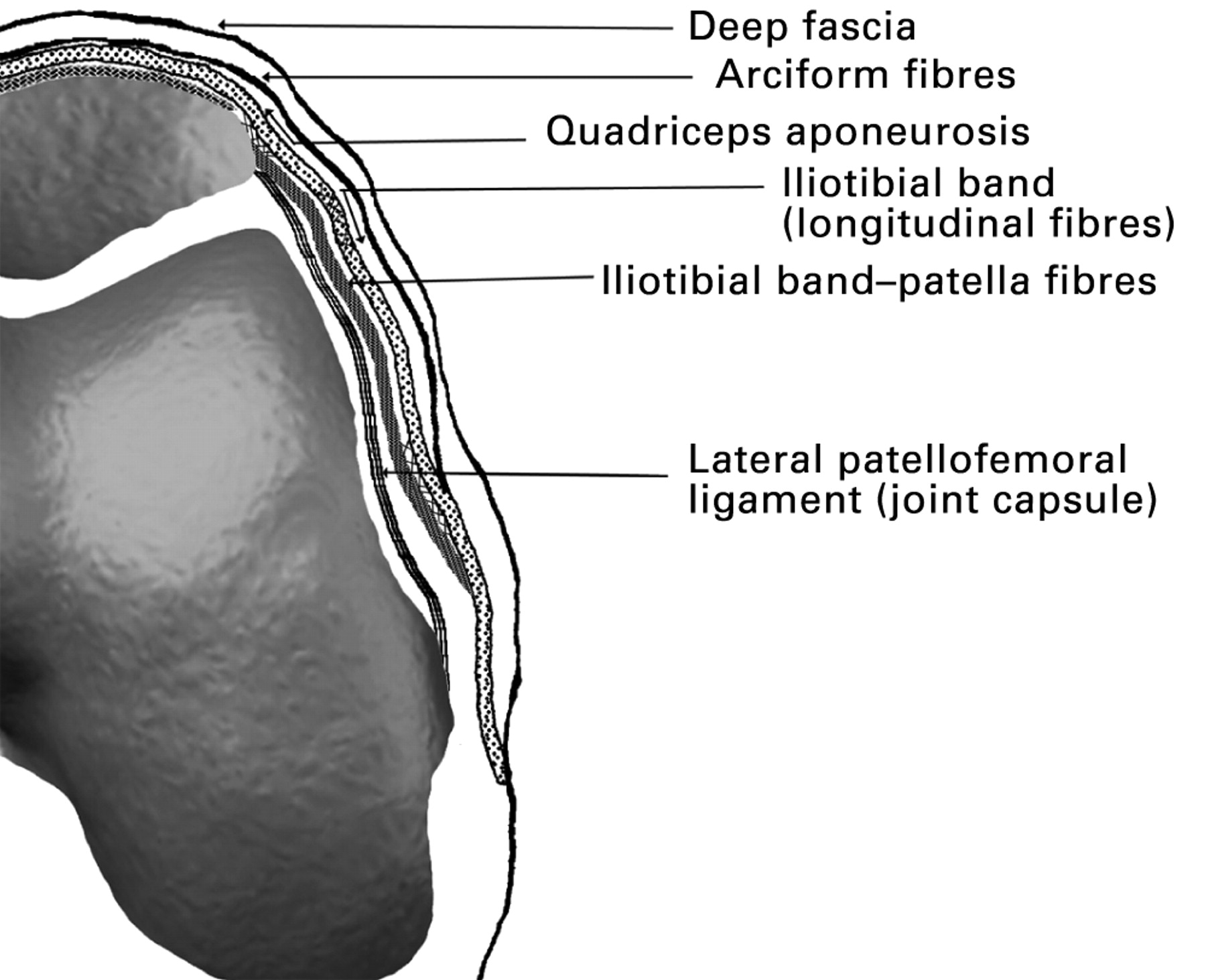
Anatomy of the Medial Retinaculum

The medial retinaculum is a thick, fibrous band of connective tissue that forms the roof of the carpal tunnel. It is located on the palmar aspect of the wrist, extending from the pisiform bone to the hook of the hamate bone. The medial retinaculum is triangular in shape, with its base attached to the pisiform bone and its apex attached to the hook of the hamate bone. It is approximately 2 cm wide and 1 cm thick.
The medial retinaculum is composed of three layers of connective tissue:
– The superficial layer is composed of dense, regular connective tissue.
– The middle layer is composed of loose, areolar connective tissue.
– The deep layer is composed of dense, irregular connective tissue.
The medial retinaculum is attached to the surrounding structures by a number of ligaments. The pisohamate ligament attaches the medial retinaculum to the pisiform bone. The hamatometacarpal ligament attaches the medial retinaculum to the hook of the hamate bone. The transverse carpal ligament attaches the medial retinaculum to the scaphoid, lunate, triquetrum, and hamate bones.
The medial retinaculum plays an important role in maintaining the integrity of the carpal tunnel. It prevents the tendons of the flexor muscles of the hand from bulging out of the carpal tunnel and compressing the median nerve. The medial retinaculum also helps to protect the median nerve from injury.
Function of the Medial Retinaculum

The medial retinaculum plays a crucial role in maintaining the integrity of the carpal tunnel and facilitating the smooth movement of flexor tendons and the median nerve.
Primary Function
The primary function of the medial retinaculum is to hold the flexor tendons in place against the carpal bones, creating a tunnel-like structure known as the carpal tunnel. This tunnel allows the tendons to glide freely during hand movements, such as flexing the fingers and wrist.
Carpal Tunnel Formation
The medial retinaculum forms the roof of the carpal tunnel, along with the transverse carpal ligament. Together, these structures create a narrow passageway through which the flexor tendons and the median nerve pass.
Implications of Thickening or Tightness
Thickening or tightness of the medial retinaculum can compress the flexor tendons and the median nerve within the carpal tunnel. This compression can lead to a condition called carpal tunnel syndrome, characterized by pain, numbness, and tingling in the hand and fingers.
Clinical Significance of the Medial Retinaculum

The medial retinaculum, a crucial structure in the wrist, plays a significant role in carpal tunnel syndrome, a common condition affecting the hand and wrist.
Carpal Tunnel Syndrome
Carpal tunnel syndrome occurs when the median nerve, which passes through the carpal tunnel, becomes compressed. The medial retinaculum forms the roof of the carpal tunnel, and its thickening or inflammation can narrow the tunnel, leading to nerve compression.
Symptoms of carpal tunnel syndrome include numbness, tingling, and pain in the thumb, index, middle, and ring fingers. These symptoms are often worse at night or after prolonged use of the hands.
Diagnosis of carpal tunnel syndrome typically involves a physical examination and nerve conduction studies to assess the median nerve’s function.
Treatment Options, Medial retinaculum
Treatment for carpal tunnel syndrome aims to reduce pressure on the median nerve. Non-surgical options include wrist splints, corticosteroid injections, and activity modification. If these measures fail to provide relief, surgery may be necessary.
Surgery involves releasing the medial retinaculum, thereby enlarging the carpal tunnel and reducing pressure on the median nerve. This procedure, known as carpal tunnel release, is generally effective in alleviating symptoms.
The medial retinaculum, a ligament that stabilizes the wrist, is crucial for tennis players like John McEnroe. His recent resurgence in the sport has shed light on the importance of this structure. Read more about John McEnroe’s latest news.
Understanding the medial retinaculum’s role in wrist stability is essential for athletes and healthcare professionals alike.
The medial retinaculum, a band of connective tissue that stabilizes the tendons in the wrist, is essential for maintaining proper hand function. Tennis legend John McEnroe, renowned for his fiery on-court demeanor, recently shared his latest news here. Despite his intense playing style, McEnroe has managed to preserve the integrity of his medial retinaculum, allowing him to continue his illustrious career on the court.
The medial retinaculum, a band of tissue in the wrist, plays a crucial role in stabilizing the carpal bones. It’s fascinating how different parts of our body work together harmoniously. Like the recent news about soft drinks recalled due to contamination, the medial retinaculum reminds us that even seemingly unrelated events can impact our well-being.
The medial retinaculum, a band of tissue in the wrist, is a pivotal structure for wrist stability and movement. Like Walt Frazier , the “Clyde” of the legendary Knicks duo, the medial retinaculum plays a dynamic role, enabling graceful hand and wrist movements, whether it’s dribbling a basketball or performing intricate piano sonatas.
Like a skilled basketball player like Bob Cousy who deftly handles the ball, the medial retinaculum acts as a protective sheath for tendons that run along the wrist. Its intricate structure ensures smooth movement and flexibility, allowing us to perform everyday tasks with ease.
As the tendons glide beneath the retinaculum, they are shielded from friction and injury, much like how Cousy’s exceptional ball-handling skills protect the ball from opponents’ attempts to steal it.